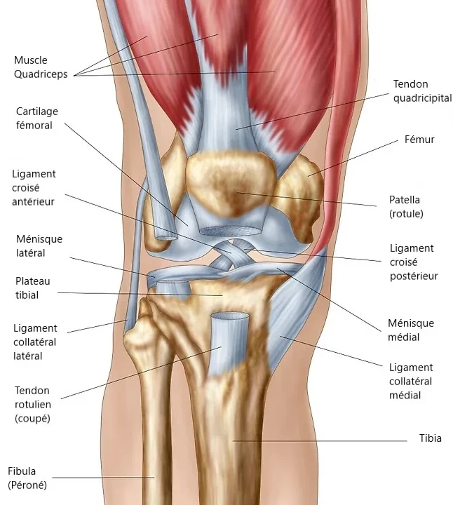
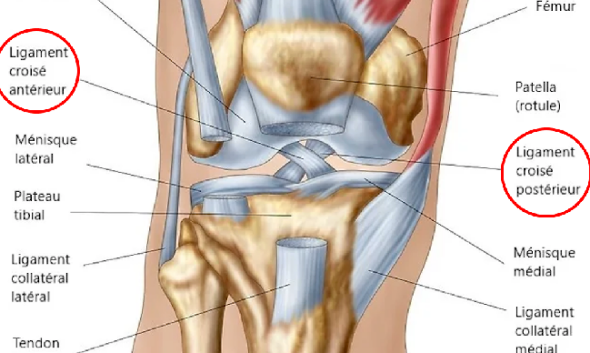
The knee refers to the joint located between the femur (thigh bone) and the tibia (shin bone). It functions roughly like a hinge, not only performing flexion/extension but also minimal rotational and sliding movements forward and backward. The distal end of the femur has the shape of two bony balls, the medial and lateral condyles, joined by a central groove, the trochlea, in which the patella (kneecap) slides during knee flexion and extension. The proximal end of the tibia is called the tibial plateau, which is a more or less flat surface. This defines three joint spaces: the medial femorotibial space, the lateral femorotibial space, and the femoropatellar space.
The articular surfaces (distal femur, proximal tibia, and articular surface of the patella) are covered with a fairly hard, whitish, very slippery substance called cartilage. This substance is constantly lubricated by a small amount of viscous fluid, synovial fluid, produced and renewed by the cells of the synovial membrane lining the inner surface of the joint. Well-lubricated cartilage allows for frictionless and painless movement between the articulating bones. After the end of growth, we all have a fixed stock of cartilage in our joints, which unfortunately cannot naturally regenerate identically.
The contact surface between the femoral condyles and the tibial plateau is increased by two crescent-shaped structures fixed to the tibial plateau, with a fairly rigid elastic consistency and a triangular cross-section, called menisci. These are the medial and lateral menisci, which cushion the contact of the femur with the tibia and also contribute to knee stability.
The joint is enveloped by a malleable but tear-resistant membrane called the joint capsule. This ensures the tightness of the joint, preventing the leakage of synovial fluid. A second circumferential envelope formed by ligaments, especially the medial collateral ligament and lateral collateral ligament, maintains the knee and prevents it from deviating into varus (bow-legged) or valgus (knock-kneed) alignment.
In the center of the joint, two powerful ligaments stabilize the knee in rotational and forward/backward translation movements. These are the anterior and posterior cruciate ligaments, which form the central pivot of the knee. Two main muscle groups move the knee: the hamstrings in the back for flexion, and the large quadriceps muscle in the front for extension. The latter is crucial for standing and walking. It is continuous with the patella and extends through the patellar tendon, which inserts on the proximal tibia—this is the knee extensor apparatus.
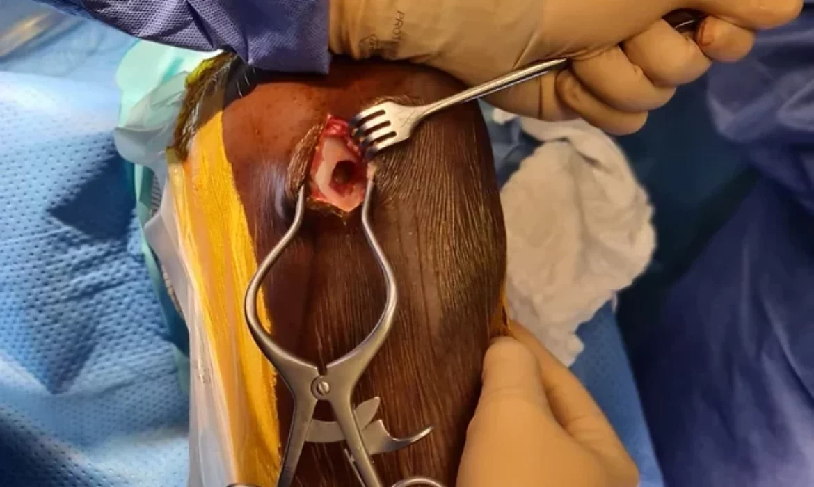
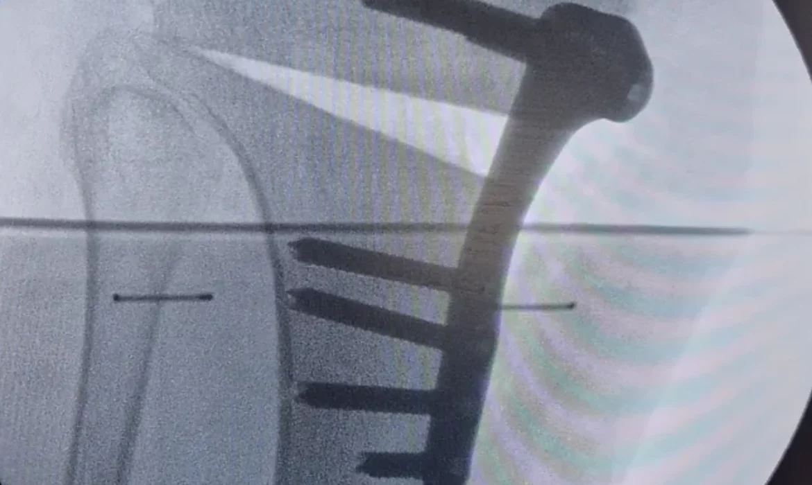
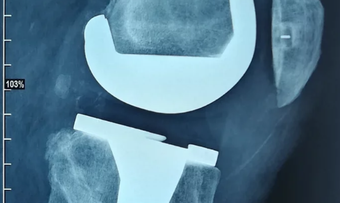
Here are the most frequently performed interventions by our team on this joint: