PLANTAR APONEUROSIS (CALCANEAL SPUR)

ANATOMY
The plantar aponeurosis, or plantar fascia, is a thick fibrous membrane located on the plantar (bottom) surface of the foot. It has a triangular shape, with its posterior part attaching to the calcaneus (heel bone) and widening towards the front of the foot to end at the metatarsal heads.
Its role is to support the arch of the foot and protect the overlying tendons.
This membrane is part of a larger fibrous complex known as the Suro-Achilleo-Plantar complex. In fact, the plantar aponeurosis is a continuation of the posterior leg fascia (Achilles aponeurosis).
Thus, this fibrous complex originates behind the knee, extends behind the leg through the Achilles tendon, reflects on the calcaneus (heel bone), and terminates at the metatarsal heads.
The plantar aponeurosis can be seen as the plantar part of a more comprehensive structure.
PATHOLOGY :
Inflammation of the plantar aponeurosis occurs due to abnormal or repetitive stresses on it. This can occur during significant physical activities such as long walks or running, as well as due to structural abnormalities in the rearfoot, whether they are static (flat feet, high arches) or dynamic (valgus, pronation, varus), leading to excessive tension on the aponeurosis.
Mechanical factors like overweight or heavy lifting can also contribute to this condition. The stress on the aponeurosis results in mechanical forces on both of its insertions: these stresses are distributed over a wide area in the front (lower pressure), but are concentrated in a very limited area in the rear, at the calcaneal insertion, where the pressures are highest.
Therefore, it is this significant tension in a limited area that causes heel pain and potential radiographic signs.
THE CLINIC :
Plantar fasciitis is the main cause of heel pain or “talalgia”. It is characterised by pain in the heel, aggravated by weight-bearing and walking, forcing patients to walk on the forefoot or the outer edge of the midfoot.
THE DIAGNOSIS :
The diagnosis is primarily clinical, based on pain elicited by palpation of the underside of the heel, often on the inner side, sometimes on the outer side.
The pain is also reproduced by tensioning the fascia, by dorsiflexion of the ankle with pressure on the metatarsal heads. Radiographic evaluation is used to look for signs of severity or chronicity, such as ossification at its calcaneal insertion (hence the name “heel spur” or LENOIR spur).
MRI and ultrasound can confirm the diagnosis by revealing inflammation of the fascia and searching for signs of severity (fissures).
The adaptation of footwear with more cushioning and a slight heel is a simple way to reduce pain at the beginning of the symptoms. Medications (pain relievers and anti-inflammatories) prescribed by your primary care physician can help combat acute pain and inflammation.
Reducing contributing factors such as rest or weight loss should be the first measures considered. Correcting structural foot problems with the use of orthotic insoles (orthopedic shoe inserts) is an excellent way to relieve inflammation of the plantar fascia.
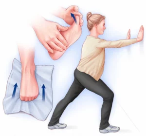
Rehabilitation physiotherapy, involving stretching of the plantar fascia and the entire suro-achilleo-plantar chain, helps reduce pressure and inflammation. Deep transverse massages can also be performed.
The use of shockwaves, mesotherapy, or local cortisone injections is excellent therapeutic methods to reduce inflammation.
Without treatment, heel pressure becomes increasingly painful, leading to limping with a lack of support and difficulties in wearing shoes.
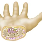
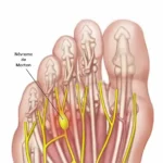
In the worst-case scenario, fibrous nodules can form within the inflamed plantar fascia, and even a rupture of the fascia can occur.
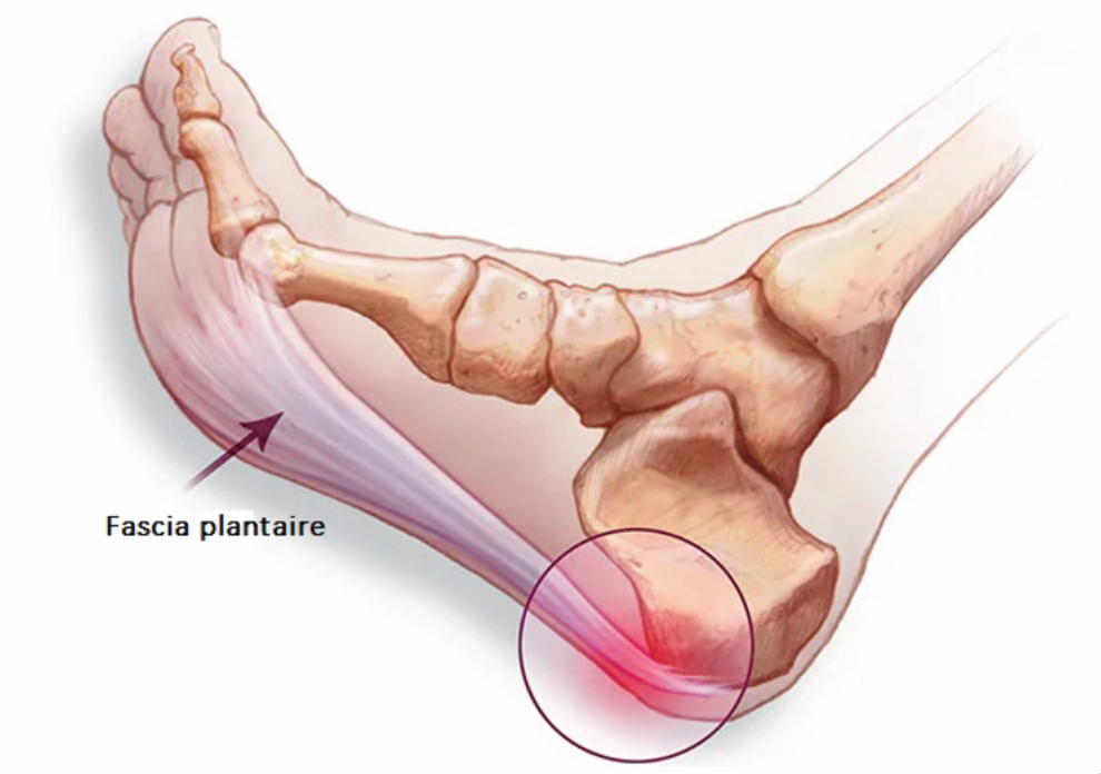
Important Point: The “ossification” image at the insertion point of the plantar fascia on the heel bone is often misinterpreted by patients as the cause of their pain, resembling a “painful spur” that should be removed.
However, this “bony spur” is not inherently painful; it is merely a reflection of the mechanical tension at the bone insertion of the plantar fascia.
Therefore, treatment involves lengthening the plantar fascia, and the removal of the ossification has no benefit!
When conservative treatment is insufficient, surgical intervention may be proposed. Surgical treatment involves partial or total sectioning of the plantar fascia, which then heals in a relaxed position, reducing pressure within it. Inflammation subsequently resolves on its own.
Sometimes, surgical treatment is part of a more comprehensive approach to correcting structural abnormalities in the midfoot or hindfoot. The procedure on the plantar fascia is just one step in releasing the midfoot or hindfoot.
HOSPITALIZATION :
Hospitalization is typically done on an outpatient basis, meaning you may go home on the same day as the surgery. However, in some cases, a one-night hospital stay may be required due to health issues or social isolation. The decision regarding the length of hospitalization will be made based on your specific health condition and needs.
THE ANESTESIA :
A preoperative consultation with an anesthesiologist is mandatory. During this consultation, the anesthesiologist will explain to you the methods and possible choices for anesthesia that are tailored to the surgery and your health condition.
During this consultation, your current medication regimen will also be reviewed. New medications may be prescribed, either before or after the surgery. The most commonly used medications include anticoagulants, antibiotics, pain relievers, and anti-inflammatories, each of which carries specific risks.
The type of anesthesia used during the surgery can be regional (affecting a specific segment of the body, from the leg to the toes), spinal (affecting the pelvis and limbs by injecting between two vertebrae), or general anesthesia.
THE TECHNIQUE:
The surgery lasts approximately 30 to 45 minutes and involves the partial or total sectioning of the plantar fascia a few centimeters before its insertion into the calcaneus. The fascia then heals spontaneously in a relaxed position over a few weeks, reducing pressure on the bone insertions, especially at the calcaneal insertion point.
The incision is made on the inner side of the heel, and its length depends on the surgical technique used.
Several surgical techniques are possible:
- “Open” surgery, which is performed under direct vision through a short incision of less than 10 cm.
- Percutaneous surgery, involving a small puncture incision of a few millimeters, allowing the sectioning of the fascia using a fine scalpel, guided by a fluoroscope (a type of X-ray machine) to monitor the instrument’s position.
- Endoscopy: This involves using a camera inserted through an incision of approximately 1 centimeter, allowing the guided sectioning of the fascia through a video image.
A tourniquet is often used to temporarily stop blood flow to the surgical area. It can be placed at the thigh, leg, or ankle level.
During the surgery, unexpected or unusual situations or events may require additional or different procedures than originally planned. After awakening and when the surgery is completed, your surgeon will explain the procedure and any additional steps that were taken.
POST-OPERATIVE CARE :
SUPPORT
Depending on your surgeon's practices and the surgical technique, you may be prescribed immobilization for a few weeks. Partial weight-bearing is usually allowed (the use of crutches is possible) during the first few weeks, depending on pain levels.
Wearing wide but closed shoes is typically resumed around the 3rd week. Returning to regular footwear usually occurs between 2 and 3 months.
POST-OPERATIVE OEDEMA
Swelling (swelling of the foot and toes) is common in foot surgery and is not a complication. Managing swelling is essential not only to alleviate pain but also to improve the quality of healing: thus, a period of rest, elevation, and the use of vein support (compression stockings or varicose stockings) can be helpful.
This swelling can last a long time (several weeks, even months) and usually does not lead to any significant issues. It may temporarily require adjusting footwear.
PAIN
Post-operative pain from this type of pathology generally poses little difficulty. Even though strong painkillers may be used in the immediate aftermath, going home with simple analgesics is the norm.
Sometimes, a more or less prolonged and complete anesthesia of the operated limb (anesthetic block or loco-regional anesthesia) is also used to decrease or eliminate the most significant pains of the first few days. A work stoppage is generally necessary after the surgical procedure. Its duration averages 6 weeks but depends on your professional activity and the surgical procedures performed.
POST-OPERATIVE CONSULTATIONS :
Your surgeon will offer you post-operative monitoring. The frequency will depend on the surgical procedure and your progress. The first consultations will focus on monitoring the healing process, the local condition and the resumption of weight-bearing.
Subsequent consultations will focus on functional recovery, sedation of pain and the return to full weight-bearing.
AND AFTER THE SURGERY ?
Even if the surgery is short and typically done on an outpatient basis, the postoperative recovery is lengthy for pain to decrease (approximately 3 to 6 months).
Indeed, this condition usually develops over many months or years before surgery is considered. Therefore, the time it takes for the inflammatory processes to decrease and for complete healing of the plantar fascia is long and occurs over several weeks.
Adapted from the information sheet of the SFCP (French Society of Foot Surgery)